Different P Wave morphology.
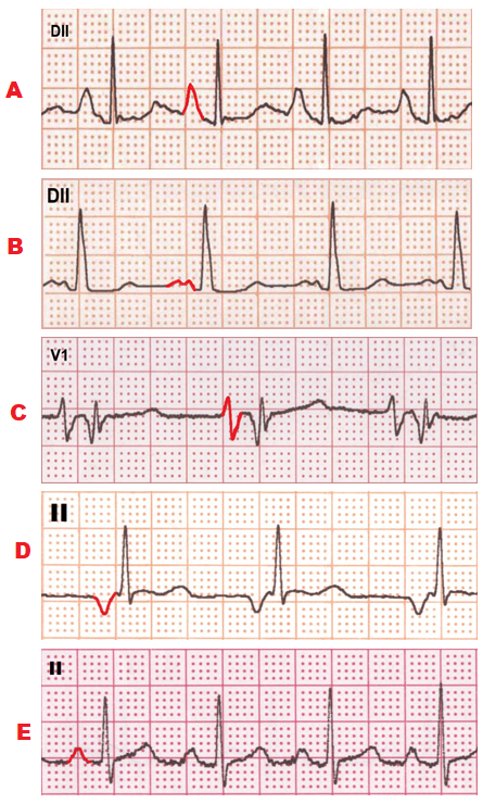
In a rhythm strip A,
presents Pulmonary P wave that
suggests right atrial enlargement. P wave with duration less than 120ms and amplitude more than 2.5mm.
In a rhythm strip B, presents Mitrale P wave that suggests left atrial enlargement. P wave with duration more than 120 ms and amplitude less than 2.5mm.
In a rhythm strip C, presents Biphasic P wave with terminal negative portion more than 40 ms and more than 1mm deep which suggests left atrial enlargement.
In a rhythm strip D, presents Inverted P wave preceding each QRS complex with normal PR interval that suggest low atrial rhythm.
In a rhythm strip E, presents Normal P wave that suggests normal sinus rhythm.


Comments
Post a Comment